This post extends on my post of last week:
How Full-Body Scanners Work – and Fail
The aim of this post is to explain x-ray backscatter scanning in further detail by examining a few of the patents that describe how x-ray backscatter full-body scanners work.
In order to receive a patent, an inventor must disclose the details of their invention in sufficient detail for a practitioner of “ordinary skill” in the relevant technical art. Thus, to understand how x-ray backscatter full-body scanners work, tracking down the patent is a good first step. Steven W. Smith received U.S. Patent 5,181,234 for this approach to full-body scanning in 1993:
A pencil beam of X-rays is scanned over the surface of the body of a person being examined. X-rays that are scattered or reflected from the subject’s body are detected by a detector. The signal produced by this scattered X-ray detector in then used to modulate an image display device to produce an image of the subject and any concealed objects carried by the subject. The detector assembly is constructed in a configuration to automatically and uniformly enhance the image edges of low atomic number (low Z) concealed objects to facilitate their detection. A storage means is provided by which previously acquired images can be compared with the present image for analyzing variances in similarities with the present image, and provides means for creating a generic representation of the body being examined while suppressing anatomical features of the subject to minimize invasion of the subject’s privacy.
The system mechanically scans an x-ray beam across the person to be examined. For each pixel, the x-ray detector collects the backscattered signal. The amount of signal collected yields the amplitude of that pixel in the processed image. Low atomic number materials (like tissue, organic materials, plastics) reflect strongly and create a bright spot. High atomic number materials like most metals reflect poorly creating a dark spot.
Minimizing radiation dose is an “important consideration:”
Radiation exposure is an important consideration in X-ray concealed object detection systems. The United States National Council on Radiation Protection (NCRP), in NCRP Report No. 91, “Recommendations on Limits for Exposure to Ionizing Radiation”, 1987, addresses this issue. In this report, the NCRP states that a radiation exposure of less than 1000 microRem per year in excess of environmental levels is negligible, and efforts are not warranted at reducing the level further. Persons employed in security facilities or frequently traveling by airlines may be subjected to many hundred security examinations per year. A yearly radiation exposure of 1000 microRem permits a single scan exposure within the range of 1 to 10 microRem for the general public. In accordance with the NCRP recommendations, radiation levels significantly higher than this present a non-trivial health risk.

The patent also provides some basic information for anyone interested in calculating dose:
In the preferred embodiment, the X-ray tube operates in the range of 35 to 70 Kilovolts with a pixel size of 12 to 120 square millimeters. The backscatter X-ray detector 17 comprises an X-ray sensitive area of 150 to 1500 square inches and is positioned 5 to 15 inches from the body of the person being examined. In another embodiment, the backscatter detector may be positioned farther from the person being examined with a corresponding increase in detector area. It has been empirically determined that these parameters are approximately optimized at the values of 50 Kilovolts, 40 square millimeters pixel area, 952 square inches X-ray sensitive area, and an 8 inch subject-to-detector distance. These technique factors simultaneously provide a CV (coefficient of variation, to be defined below) in the range of 2 to 10 percent and X-ray dose in the range of 1 to 5 microRem, as shown by the following analysis of a preferred embodiment of the present invention….

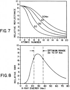
In the preferred embodiment, The optimum x-ray energy is about 30keV. The “reflectance slope” is steepest at this energy level for backscattering around atomic number Z = 6 (Carbon) to Z = 8 (Oxygen), thus providing maximal contrast. Keep in mind that this patent is nearly twenty years old, and the state of the art may have changed. Specific parameters of the embodiment preferred in 1991 may no longer hold true today. Still, the information in U.S. Patent 5,181,234 provides a useful starting point.
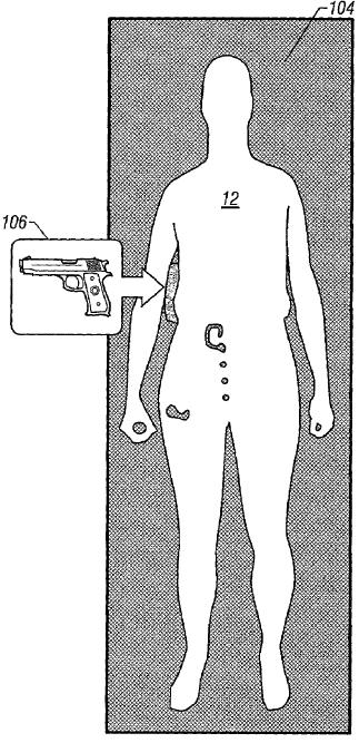
A wide variety of follow-on inventions improve on the basic scheme. One problem area is detection of contraband hidden along the edge of a body. A typical backdrop returns little reflection, making it possible to hide a low reflection object, like a gun. U.S. Patent 7,110,493 addresses this issue by placing a low atomic number backdrop behind the person being examined to reflect back additional x-rays. This helps make the background bright enough to contrast metallic objects hidden along the side of a body.
Want to learn more? A good place to start is by reviewing the 63 issued U.S. patents that cite U.S. 5,181,284 as prior art. When I get a chance, I’ll dig deeper and report further. If you are impatient, feel free to dive in and report back.


2 thoughts on “How X-Ray Backscatter Full-Body Scanners Work”